MEDICALConverting a DICOM file to a 3D Model
Medical
- Introduction of Medical Materials
- Quality Control System
- ORIGINAL PRODUCTS
- Applications
- CASE STUDIES
Converting a DICOM file to a 3D Model
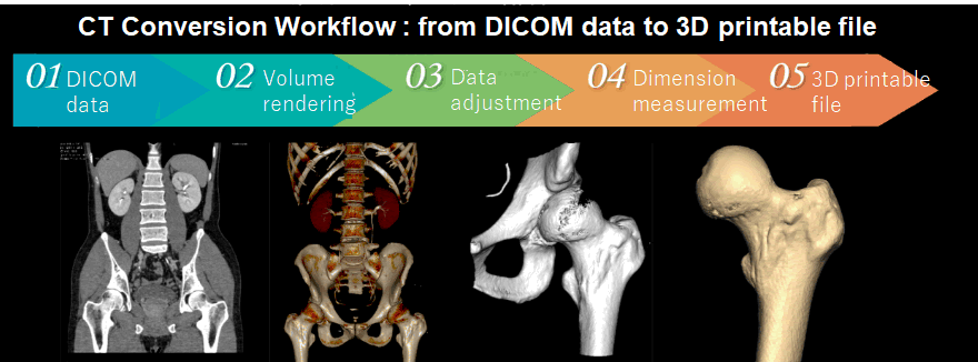
Digital Imaging and Communications in Medicine (DICOM) and is a standard for handling, storing, printing, and transmitting information in medical imaging. A DICOM file is a series of images generated from a CT scan and consists of many hundreds of individual cross-sections of an object. The combination of all of these 2D images creates a 3D volume which can be processed into a DICOM file suitable for 3D printing.
Yasojima can process and manipulate DICOM data and files in order to produce 3D printed models with 3D printing or additive manufacturing.
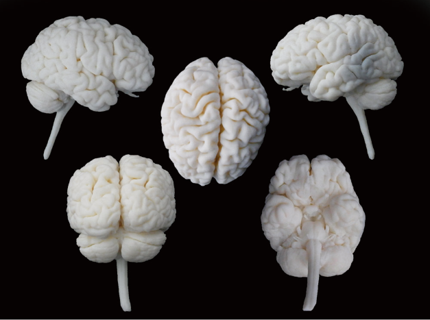
DICOM Data Extraction and Modification of a Brain
The attached membrane and the difference in the threshold value of the brain with above and below make it very difficult to extract the brain data. 3D brain data files are large in size complex, processes have to be taken to have a complete 3D data for modeling.
①DICOM reading and volume rendering. Remove unnecessary objects from the 3D data.

②Data correction, noise reduction, and dimensional design.
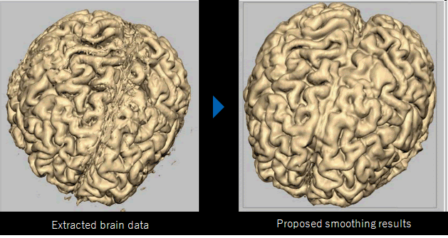
③The 3D brain data is complete.
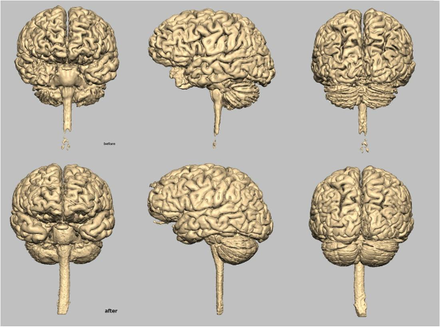
④Data is 3D printed to replicate a realistic anatomical model of the original 3D brain data for use in surgical training, simulation, or demonstration.
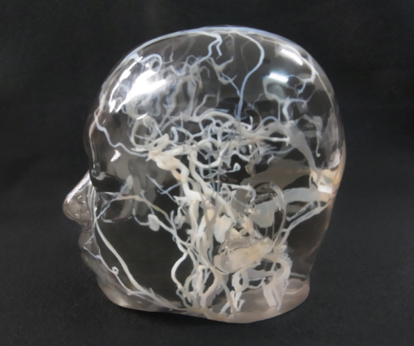
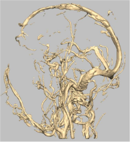
Blood Vessel Data Extraction and Modification
As with the brain, it is difficult to extract 3D data of blood vessels all at once due to the difference in threshold values, the data needs to be collected little by little while controlling extracting values.
①Extract data of blood vessels
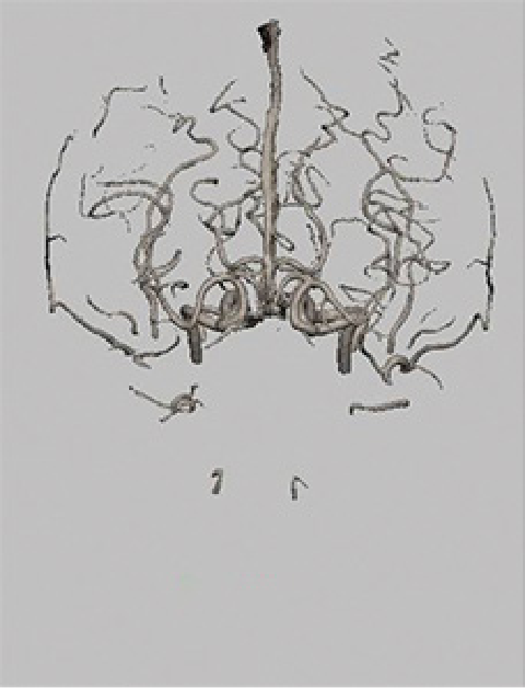
②Extract necessary data while controlling threshold and the numerical value to be extracted.
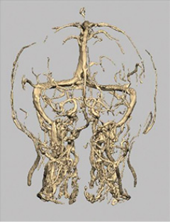
③Separate arteries and veins, and perform volume rendering.
arteries
veins
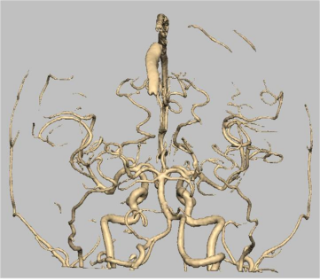
④Synthesize the coordinates of arteries and veins based on their structures and functions of blood vessels.
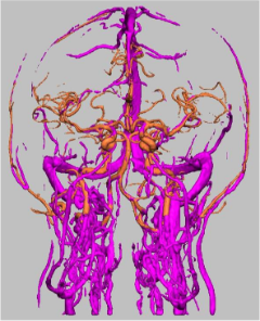
⑤3D print a visualization model with the completed 3D blood vessel data